
Traumatic diaphragmatic hernia
Dr. Dominique Penninck
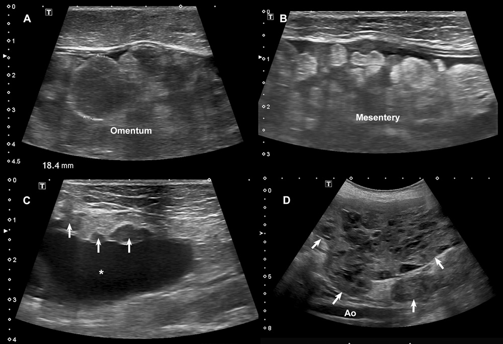 Figure 15.17. Carcinomatosis and sarcomatosis. A and B: In these two cats with pancreatic adenocarcinoma, nodules of variable echogenicity are disseminated in the omentum and mesentery. C: In one of these cats (same as in A), hypoechoic nodules also invade the peritoneal lining and adjacent abdominal wall. D: In this dog with undifferentiated soft tissue sarcoma, masses were found throughout the abdominal cavity, consistent with sarcomatosis. co-authored by Marc-André d'Anjou and Éric Norman Carmel
Questions or comments (2)
Parmjit Dhillon
This is good stuff for learning and refreshing. You guys are doing great by sharing these clips and images and pricing the quality of veterinary medicine globally. Keep but up. Thanks
Parm
Marc-Andre Author
Thank you for these words. :-) As you pointed out, the objective here is to level up the expertise of small animal ultrasound across the globe. And having vets like you sharing their thoughts and experience will only benefit to everyone, starting with Dominique and I!
Sign in to ask a question or leave a comment.